University of Washington is the Home of Practical 3D Printing
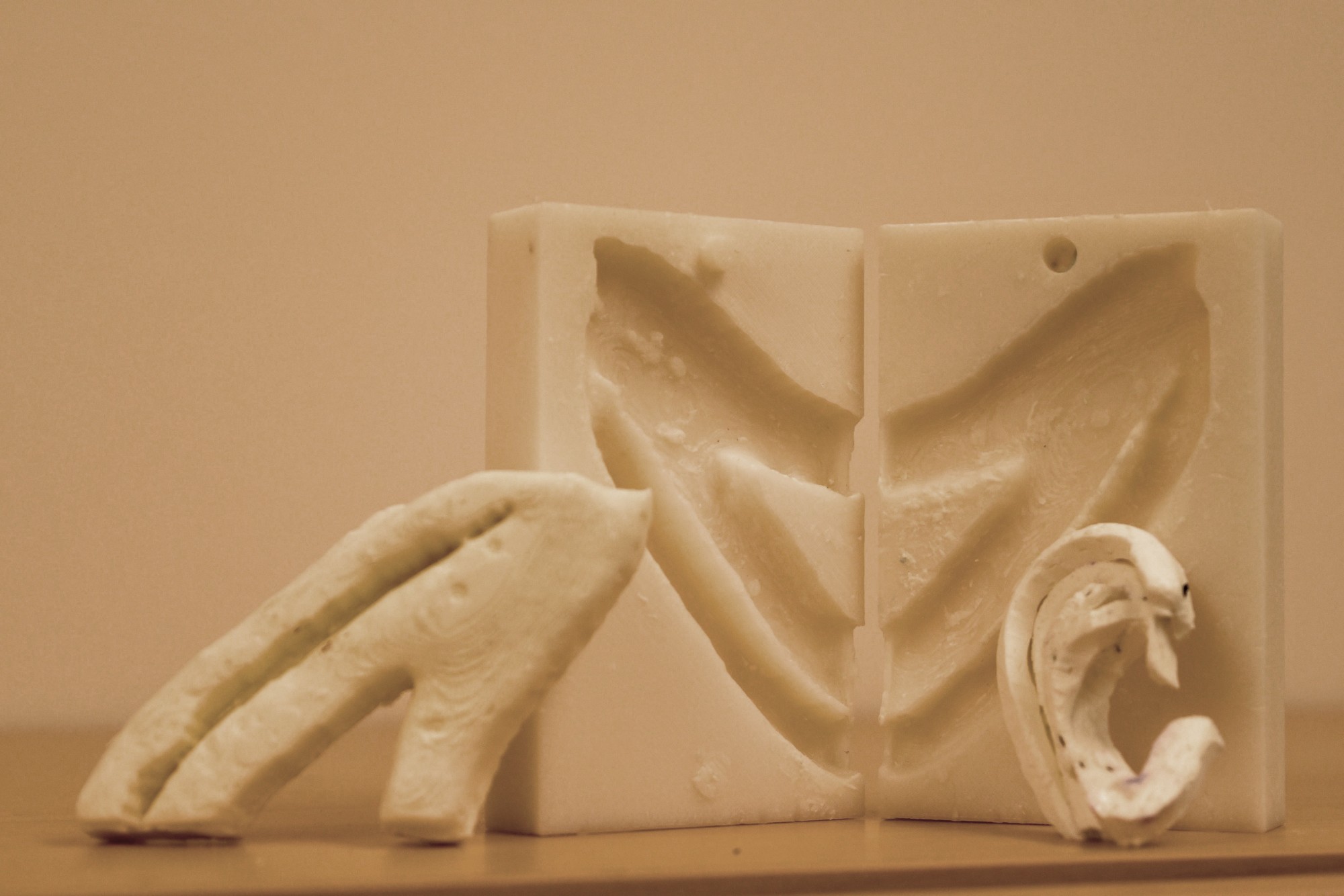
An example of a practice rib (and practice ear) made from CT scan data and a 3D printed mold. Courtesy of UW.


Latest News
October 1, 2015
The adoption of additive manufacturing (AM) and rapid prototyping encourages people to fail big and fail often because the costs of doing so are relatively small. That mindset is great for innovation, but can occasionally lead to ideas that will never be practical.
The culture surrounding 3D printing at the University of Washington (UW) appears to have different ideas about how to best leverage the technology. The students and researchers at UW still think big, but seem to be able to remain practical about what is currently possible with AM. This attitude leads to projects that are viable right now, rather than projects that may become viable 20 years down the road.
 An example of a practice rib (and practice ear) made from CT scan data and a 3D printed mold. Courtesy of UW.
The latest example of this approach is the combination of 3D printing and replacement ears. Other research facilities have gone all in with attempting to print “bionic” ears, while UW has taken a more nuanced approach to the same goal. Rather than attempting to print entire ears, Angelique Berens and her fellow researchers have chosen to assist doctors who are recreating ears.
Generally, to treat children with underdeveloped or missing ears, surgeons borrow some of the patient’s rib cartilage and then carve it into the shape of an ear. The problem is that most doctors don’t have a lot of practice carving ears, and it’s difficult to find a material similar in feel to the cartilage used for the procedure.
“It’s a surgery that more people could do, but this is often the single biggest roadblock,” said Kathleen Sie, a UW Medicine professor of otolaryngology and director of the Childhood Communication Center at Seattle Children’s. “They’re hesitant to start because they’ve never carved an ear before. As many potatoes and apples as I’ve carved, it’s still not the same.”
The solution discovered by the research team was to use CT scan data to create a 3D printed mold of a patient’s ribs. A special silicone solution is then poured into the mold and allowed to set. The finished product is an artificial rib that, according to the UW team, feels and behaves more like actual rib cartilage than anything else thus far.
“It’s a huge advantage over what we’re using today,” said lead author Angelique Berens, a UW School of Medicine otolaryngology surgery resident. “(Today) you literally take a bar of Lever 2000 (soap) while the attending is operating and you carve ear cartilage. It does teach you how to get the shape right, but the properties are not super accurate — you can’t bend it, and sewing it is not very lifelike.”
Below you’ll find a video about 3D printing at UW.
Source: UW
Subscribe to our FREE magazine, FREE email newsletters or both!
Latest News
About the Author
John NewmanJohn Newman is a Digital Engineering contributor who focuses on 3D printing. Contact him via [email protected] and read his posts on Rapid Ready Technology.
Follow DE